How HP-OCT™ harnesses the power of beamlets
Cylite’s HP-OCT™ shares several similarities with existing spectral-domain OCTs (SD-OCT). What sets it apart though is the way it measures the eye. While conventional SD-OCTs rely on a single scanning measurement point, HP-OCT™ measures the eye using 1,008 simultaneous measurement points, or ‘beamlets’.
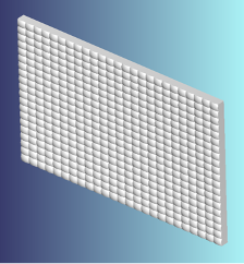
The novel HP-OCT™ was designed using micro-optics technology, rather than the fibre optics technology used in conventional SD-OCT systems. Within the micro-optics technology is the inclusion of a micro-lens array (MLA), utilised to split the light source into 1,008 individual ‘beamlets’.
A micro-lens array (MLA) is, as the name suggests, an array of miniature optical lenses (also called lenslets) arranged in a regular pattern. The MLA used in the HP-OCT™ has a 24 x 42 array of lenslets to give a total of 1,008 lenslets. By optimising the optical design, the MLA can then split the light source within the HP-OCT™ into 1,008 individual parallel beams of light or ‘beamlets’. This unique optical design ensures distortion-free, telecentric projection of the beamlets onto the eye.
The bespoke internal micro-optics design of the HP-OCT™ therefore enables simultaneous A-scans to be measured in single ‘snapshot’ capture, which we term a ‘frame’. Each frame consists of 1,008 A-scans and acquires 300 frames a second, resulting in an A-scan rate of 302,400 A-scans per second.
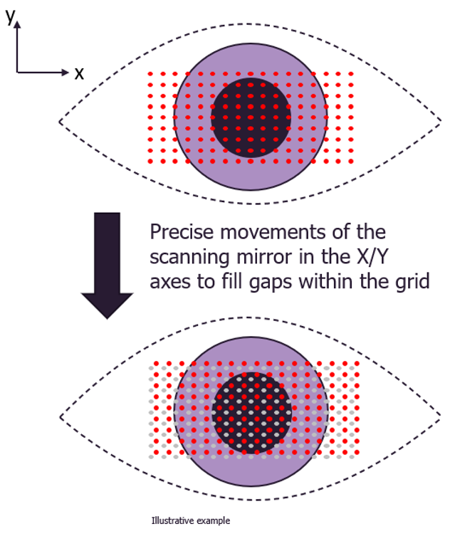
Each frame contains a full 3D volume image of the region of interest (i.e. anterior segment or retina). It is this unique beamlet ‘snapshot’ capture that overcomes the natural eye motion constraints, or motion artefacts.
To further enhance the quality of the images and provide dense coverage between each point, the position of the beamlet array on the eye is moved very slightly between each frame in a precise, predetermined pattern. This motion is made possible by the micro-optics technology within the HP-OCT™, which allows all 1,008 beamlets to scan both the X and Y axes simultaneously, thus allowing the device to acquire dense and distortion-free volumetric data.
HP-OCT™ – Redefine your perspective
- Insensitive to movement for clinical applications due to ‘snapshot’ capture.
- Accurate anterior and posterior corneal topography
- Complete optical biometry
- Full anterior chamber OCT volumetric imaging
- High speed retinal OCT volumetric imaging
- >300,000 A-scans per second